Table of Contents >> Show >> Hide
- Why a CT scan is so important in kidney cancer
- What a CT scan can show when kidney cancer is suspected
- What a CT scan cannot tell with complete certainty
- Why contrast dye is often part of the test
- What to expect before the CT scan
- What happens during the CT scan
- What to expect after the scan
- How CT fits with other kidney cancer tests
- Questions to ask your doctor after the scan
- Patient experiences: what this process often feels like in real life
- Final takeaway
Hearing the words “You need a CT scan” can make your brain do acrobatics. One minute you are thinking about lunch, and the next you are wondering whether a machine shaped like a giant donut is about to reveal something life-changing. If your doctor suspects kidney cancer, a CT scan is often one of the most important imaging tests in the process. It helps doctors look closely at the kidneys, nearby blood vessels, lymph nodes, and sometimes the chest, abdomen, and pelvis to understand what is going on.
The good news is that a CT scan is fast, common, and usually much less dramatic than people imagine. The harder part is often the waiting, not the scan itself. In this guide, we will break down what a CT scan for kidney cancer shows, why contrast dye is often used, what the test can and cannot tell your doctor, and what you can realistically expect before, during, and after the appointment.
Why a CT scan is so important in kidney cancer
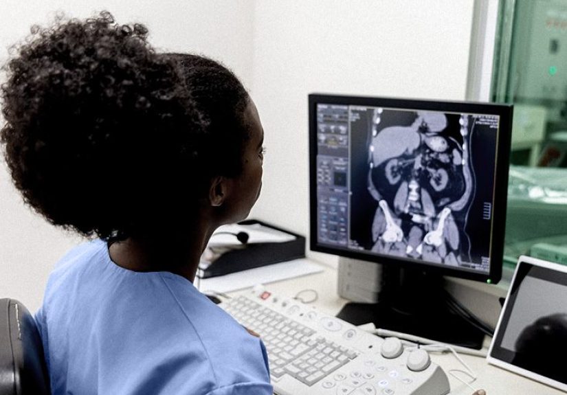
A CT scan for kidney cancer uses X-rays and computer technology to create detailed cross-sectional images of the body. Think of it as a way to look at the kidney in thin layers instead of one flat snapshot. That matters because kidney tumors can be sneaky. Some are found only after symptoms develop, but many are discovered by accident during imaging done for something else, such as abdominal pain, kidney stones, or blood in the urine.
When doctors suspect a kidney mass, CT is often the workhorse imaging test because it can show detail quickly and clearly. It helps answer several big questions at once: Is there a mass in the kidney? How large is it? Does it look solid or cystic? Has it spread into nearby tissue, lymph nodes, or blood vessels? Are there signs the disease has traveled to places like the lungs?
That is a lot of responsibility for one scan, but CT is built for exactly this kind of detective work. It is especially useful for mapping the anatomy before treatment, whether the next step is surveillance, biopsy, surgery, or another approach.
What a CT scan can show when kidney cancer is suspected
1. Whether there is a mass in the kidney
The first job of the scan is simple in theory and emotionally loaded in real life: find out whether there is a lesion or mass in the kidney. A CT scan can often detect tumors that are too small to feel on an exam and too quiet to cause obvious symptoms.
It can also help distinguish between a simple cyst and a more concerning solid mass. That distinction matters because not every kidney mass is cancer. Some are benign. Others need closer evaluation. The scan helps the radiologist describe what the mass looks like and whether it behaves in a way that raises suspicion for renal cell carcinoma, the most common type of kidney cancer.
2. The size, shape, and exact location of the tumor
If there is a tumor, the CT scan can show its size, shape, and location. That may sound like dry technical language, but it directly affects treatment decisions. A small tumor tucked away at the edge of the kidney may be approached very differently from a larger tumor near the center, where blood vessels and drainage structures live.
Surgeons use this information to decide whether a partial nephrectomy may be possible, meaning removal of just the tumor and a margin of healthy tissue, or whether a radical nephrectomy, removing the whole kidney, might be more appropriate.
3. Whether the mass enhances with contrast
Many CT scans done for possible kidney cancer use IV contrast. Contrast helps doctors see how the mass behaves after the dye circulates through the bloodstream. This is important because suspicious kidney tumors often enhance, meaning they light up differently from surrounding tissue once contrast is given.
That does not mean contrast turns the scan into a crystal ball. It does mean it gives radiologists a much better view of the tumor’s blood supply and internal character. In many cases, this helps separate a harmless fluid-filled cyst from a mass that deserves serious attention.
4. Whether nearby veins or tissues are involved
Kidney cancer is not always content to stay in its lane. A CT scan can help show whether a tumor has extended into the fat around the kidney, the adrenal gland, or nearby veins. In some cases, kidney tumors can grow into the renal vein and even toward the inferior vena cava, the large vein that carries blood back to the heart.
That information is crucial for staging and surgical planning. It tells the care team whether the disease appears confined to the kidney or whether it has become more locally advanced.
5. Whether cancer may have spread
A CT scan is also used to look for metastatic disease, which means cancer has spread beyond the kidney. Depending on the situation, doctors may order CT imaging of the abdomen and pelvis, and sometimes the chest as well. The lungs are a common place kidney cancer can spread, so chest imaging often becomes part of the bigger picture.
If enlarged lymph nodes or suspicious lesions appear elsewhere, the scan helps guide the next conversation. That may involve more imaging, a biopsy, or a different treatment plan than what would be used for cancer limited to the kidney.
What a CT scan cannot tell with complete certainty
As useful as CT is, it is not magic. It can strongly suggest kidney cancer, but it does not always provide a definitive answer on its own. Some kidney masses that look suspicious on imaging turn out to be benign. Others may require a biopsy or surgical removal before the pathology report settles the issue.
That is why your doctor may talk about the scan in terms such as “suspicious for malignancy” rather than saying, “This absolutely is cancer,” based on imaging alone. In other words, CT is often the star witness, but pathology is still the judge and jury.
Why contrast dye is often part of the test
For many people being evaluated for kidney cancer, the scan is done with iodinated IV contrast. The dye helps important structures stand out more clearly, including blood vessels and the tumor itself. In some cases, oral contrast may also be used, especially if doctors want a clearer view of abdominal structures around the kidneys.
Before contrast is given, your team may check your kidney function with blood work. This is especially common if you have kidney disease, diabetes, high blood pressure, dehydration, or other risk factors. If your kidneys are not working well or you have had a serious contrast reaction in the past, your doctor may recommend a different imaging strategy, such as MRI or ultrasound.
You should also tell your care team if you take metformin, have had contrast allergies before, or might be pregnant. None of that is “extra information.” It is exactly the kind of information the imaging team wants before they press the big shiny button.
What to expect before the CT scan
Getting ready at home
Your instructions may vary depending on whether contrast is planned, but many people are asked not to eat for a few hours before the scan. You may still be allowed to drink clear liquids and take routine medications, depending on your doctor’s directions.
Wear comfortable clothing, but be prepared to change into a gown. Leave jewelry and metal accessories at home if possible. This is not a fashion critique. Metal can interfere with imaging, and nobody wants a belt buckle stealing the spotlight from your kidneys.
Questions the staff may ask
Expect questions about:
- Medication allergies
- Prior reactions to contrast dye
- Kidney disease or reduced kidney function
- Diabetes and metformin use
- Pregnancy status
- Recent illness or dehydration
These questions are routine and designed to keep the scan safe, not to interrogate you like a medical game show contestant.
What happens during the CT scan
When you arrive, a technologist may place an IV line if contrast is being used. You will lie on a narrow table that slides through the scanner. Unlike an MRI, the CT scanner is not usually a long tunnel. It is more like a short ring or donut, which is one reason many people find it easier to tolerate.
The technologist will move to a nearby control room but can still see and talk with you. During the scan, you may be asked to hold your breath for a few seconds at a time. Staying still helps keep the images sharp.
If you receive IV contrast, you may notice a warm flushing sensation, a brief metallic taste in your mouth, or the strange but classic feeling that you might have just wet your pants even though you did not. That sensation is common, short-lived, and one of the least glamorous but most memorable parts of the test.
The scan itself is usually quick. The imaging portion may take only a few minutes, while the total visit may be under 30 minutes depending on preparation and contrast timing.
What to expect after the scan
Most people can go back to normal activities soon after the test unless they were told otherwise. If you had contrast, you may be encouraged to drink fluids afterward. Some people feel completely normal right away. Others may feel a little tired, mostly from stress, fasting, or the emotional buildup rather than the scan itself.
Call your healthcare team if you develop symptoms such as rash, worsening swelling at the IV site, trouble breathing, or anything that feels significantly off after you leave. Serious reactions are uncommon, but “probably fine” is not a diagnosis.
How CT fits with other kidney cancer tests
A CT scan is important, but it is rarely the whole story by itself. Your doctor may also use:
- Urine tests to look for blood or other clues
- Blood tests to evaluate kidney function, blood counts, and overall health
- Ultrasound to help tell whether a mass is solid or fluid-filled
- MRI if CT contrast is not a good option or if more detail is needed
- Biopsy in selected cases to confirm the diagnosis
In some cases, especially with smaller masses, the care team may even recommend active surveillance rather than immediate treatment. That means careful follow-up imaging over time to watch how the mass behaves. So yes, a CT scan may launch the treatment plan, but it may also launch a period of careful observation instead of instant action.
Questions to ask your doctor after the scan
Once the scan is done, the next challenge is usually understanding the report without accidentally convincing yourself that every unfamiliar term means doom. Ask questions such as:
- Was a mass seen, and if so, how large is it?
- Does it look more like a cyst, a benign tumor, or a possible cancer?
- Is there any sign it has spread beyond the kidney?
- Do I need more imaging, a biopsy, or a referral to a urologist?
- Would active surveillance, surgery, or another treatment be the likely next step?
Bring someone with you if possible, or take notes. Medical conversations move fast, especially when your own nervous system is busy yelling, “Please translate all of this into normal human English.”
Patient experiences: what this process often feels like in real life
On paper, a CT scan for kidney cancer sounds clean and clinical. In real life, it often begins in a much messier way. Someone gets a scan for persistent back pain, kidney stones, a urinary issue, or blood in the urine. They expect one answer and get a surprise sentence instead: “There is a mass on the kidney.” That moment can feel like the floor shifted two inches to the left.
Many people say the hardest part is not the scan itself. It is the stretch between the order, the appointment, and the final explanation from a doctor who knows how to interpret the images in context. During that window, normal life continues in a very rude way. Emails still arrive. Laundry still exists. The dog still wants dinner. Meanwhile, your brain has started auditioning worst-case scenarios.
The scan day is usually calmer than expected. Patients often describe the imaging suite as efficient, quiet, and surprisingly ordinary. There is paperwork, a few safety questions, and then the gown. Nobody enjoys the gown, but it is part of the ritual. If contrast is used, the IV placement may be the most physically uncomfortable part for some people. After that, most are struck by how fast the process moves.
During the scan, the most common reactions people mention are the sudden warmth from the contrast, the metallic taste, and a strange sense that something dramatic should be happening because the situation feels big, even though the machine is just doing its job with professional boredom. The technologist may ask you to hold your breath, stay still, and breathe again. Then, almost suspiciously quickly, it is over.
What follows is often a different kind of experience: waiting for the report, reading too much into online terms, and trying not to become an honorary radiologist by midnight. Some people see words like enhancing mass, indeterminate lesion, or suspicious renal neoplasm and immediately panic. Others cling to every phrase that sounds less alarming, such as small or confined to the kidney. Both reactions are very human.
There is also the emotional roller coaster of uncertainty. Some patients learn that the mass is likely cancer and move quickly toward surgery. Others find out that it may be a benign lesion, a complex cyst, or a small tumor suitable for surveillance. Some need an MRI or biopsy before the path becomes clear. This is why the CT scan matters so much: it narrows the mystery, even when it does not completely end it.
Another common experience is relief mixed with fear. Relief that something was found before symptoms became severe. Fear of what comes next. For many patients, the CT scan is the first real turning point. It changes the conversation from vague concern to specific planning.
If that is where you are right now, it helps to remember that a CT scan is not the end of the story. It is the map. It gives your care team a clearer picture of what they are dealing with so decisions can be based on evidence instead of guesswork. And while the emotional side of the process is real, the practical side is often manageable one step at a time: get the scan, get the results explained, ask questions, and move to the next decision with as much clarity as possible.
Final takeaway
A CT scan for kidney cancer is one of the most useful tools doctors have for finding a kidney mass, evaluating how suspicious it looks, checking whether it has spread, and planning what happens next. It often shows the size, location, and behavior of a tumor in remarkable detail, especially when contrast is used. Just as important, it helps rule out certain concerns and narrow the field when the diagnosis is still uncertain.
For patients, the experience is usually faster and less physically uncomfortable than expected. The emotional side is the bigger challenge. But understanding what the scan can show, why it is ordered, and what happens during the appointment can make the process feel less like a mystery and more like a step toward answers. And when it comes to possible kidney cancer, answers are powerful.