Table of Contents >> Show >> Hide
- Quick Anatomy: Two “Naviculars,” One Big Misunderstanding
- How These Fractures Happen
- Symptoms That Make Clinicians Suspicious
- Diagnosis: Why X-Rays Aren’t Always the Hero
- Treatment Options: What Usually Happens Next
- Healing Timelines and Return to Activity
- Complications to Know: Nonunion and Blood Supply Drama
- Rehab Basics: What Therapy Often Focuses On
- When to Seek Care Fast
- 5 Practical Tips to Avoid a “Hidden” Navicular Fracture (and Delays)
- Experiences: What Recovery Can Feel Like (and What People Wish They Knew)
- Conclusion
“Navicular fracture” sounds like one specific injury, but it’s actually a two-for-one confusion special.
In the foot, the navicular is a midfoot bone that helps form your arch. In the wrist,
“navicular” is an older name many people still use for the scaphoid, a small carpal bone near the base
of your thumb. Different bones, different biomechanics, different casts, same theme: these fractures can be sneaky,
and delays can make healing harder than it needs to be.
This guide breaks down navicular fractures in the foot and “navicular” (scaphoid) fractures in the wrist in plain,
standard American Englishplus practical examples, what diagnosis usually looks like, typical treatment paths, and what
recovery often feels like in real life. (And yes, we’ll keep it humanno medical robot voice.)
Quick Anatomy: Two “Naviculars,” One Big Misunderstanding
The foot navicular (tarsal navicular)
The tarsal navicular sits in the midfoot, roughly at the top/inside of your arch. It acts like a connector
between the rearfoot and midfoot and helps distribute forces when you walk, run, jump, andlet’s be honestsprint to catch
an elevator that is definitely not waiting.
The wrist “navicular” (scaphoid)
In wrist-land, “navicular fracture” usually means a scaphoid fracture. The scaphoid sits on the thumb side
of the wrist and helps coordinate motion between carpal bones. It’s also famous for being dramatic about healing,
partly because its blood supply can be limitedespecially toward the proximal (forearm-side) end.
How These Fractures Happen
Foot navicular fractures: trauma vs. stress
Foot navicular fractures generally show up in two main ways:
-
Acute traumatic fractures: a sudden force (like a fall, car crash, or a hard twist) causes a break.
These can involve the navicular body or small “chip” fragments where ligaments attach. -
Navicular stress fractures: repetitive loading (often running, sprinting, or jumping sports) creates tiny
cracks over time. This is a “high-risk” stress fracture in sports medicine because nonunion can happen if it’s missed or
pushed through.
A classic scenario: a competitive runner ramps up mileage fast, switches to a harder surface, starts feeling vague midfoot
pain, and assumes it’s “just tight shoes.” Weeks later, they’re limping and bargaining with their foot like it’s a coworker
who refuses to do their part of the project.
Scaphoid (wrist/navicular) fractures: the FOOSH story
The most common scaphoid-fracture origin story is a fall on an outstretched hand (often abbreviated FOOSH).
Think: slipping on wet tile, catching yourself during basketball, or wiping out on a bike. The wrist extends, the scaphoid
takes the load, and the bone can cracksometimes with surprisingly mild swelling at first.
Symptoms That Make Clinicians Suspicious
Foot navicular fracture symptoms
- Dorsal midfoot pain (pain on the top of the foot), often hard to pinpoint at first
- Pain with running/jumping that improves with rest, then returns when activity resumes
- Swelling or tenderness over the midfoot/arch region
- Limping or pain with push-off (toe-off) during walking
Stress fractures can be especially vague early onmore of an annoying “ache” than a dramatic “I broke my foot” moment.
That vagueness is exactly why they get ignored.
Scaphoid (wrist) fracture symptoms
- Pain near the base of the thumb on the wrist side
- Swelling and tendernessoften in the anatomic snuffbox (that small hollow when you extend your thumb)
- Pain with pinching, gripping, pushing, or pulling (opening jars becomes a villain)
- Sometimes the wrist feels “sprained,” but it doesn’t improve like a typical sprain
Diagnosis: Why X-Rays Aren’t Always the Hero
Here’s the frustrating truth: both navicular stress fractures in the foot and scaphoid fractures in the wrist can be
missed on early X-rays. That doesn’t mean anyone is incompetentit means bone injuries don’t always show
their hand immediately.
Foot navicular imaging
Clinicians usually start with plain radiographs (X-rays). If suspicion stays high (especially in athletes
with persistent midfoot pain), CT or MRI is often used to confirm the fracture and define
the pattern. CT is particularly useful for seeing bony detail; MRI can pick up stress injury earlier because it also shows
bone marrow edema (inflammation in the bone).
Practical example: if a runner has midfoot pain for weeks, a normal X-ray does not automatically mean “you’re fine.” It may
mean “we need the right camera lens.”
Scaphoid (wrist) imaging
A suspected scaphoid fracture typically starts with X-rays (including specialized scaphoid views). If the X-ray is negative
but physical exam findings strongly suggest a fracture (especially snuffbox tenderness), many clinicians treat it as a fracture
anywayimmobilizeand arrange follow-up imaging or advanced imaging such as MRI or CT. This “assume it until proven otherwise”
approach exists because delayed diagnosis increases the risk of nonunion and long-term wrist issues.
Treatment Options: What Usually Happens Next
Treatment depends on fracture location, displacement (whether bone fragments shifted),
stability, and your goals (for example, an elite athlete’s timeline may differ from someone who just wants to
type without wincing).
Foot navicular fracture treatment
For many navicular stress fractures and some nondisplaced traumatic fractures, the initial plan is usually:
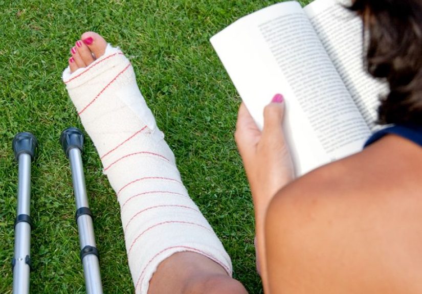
- Immobilization in a cast or boot
- Non-weightbearing (often with crutches) for a period recommended by the treating clinician
- Activity modification (translation: no “testing it” with a quick run)
- Follow-up imaging if needed to confirm healing
Surgery may be considered if there’s displacement, joint surface involvement, multiple fragments, high-risk stress fracture
patterns, nonunion, or if a faster/more predictable union is needed for certain high-demand cases. Surgical options can involve
screws and/or bone grafting depending on the situation.
Scaphoid (wrist/navicular) fracture treatment
Nondisplaced scaphoid fractures are often treated with castingcommonly a thumb spica style
castbecause immobilizing the thumb helps reduce stress across the scaphoid during healing. Healing time varies based on where the
fracture is (distal, waist, proximal), because the more proximal the fracture, the more likely blood supply is compromised.
Surgery is more likely when the fracture is displaced, unstable, involves the proximal pole, or when nonunion
risk is high. In athletes or people who need faster return to function, surgical fixation may also be discussedalways balancing
benefits, risks, and your actual life (jobs, caregiving, sports seasons, and the very real fact that casts don’t fold laundry).
Healing Timelines and Return to Activity
Timelines vary, so think of these as the “ballpark” your clinician will refine based on imaging, symptoms, and exam findings.
Foot navicular
Navicular stress fractures often require a meaningful rest periodcommonly weeks of immobilization and restricted weightbearing
before gradually returning to walking, then strengthening, then running. Return-to-sport is typically staged: pain-free walking,
then low-impact conditioning, then controlled impact, then sport-specific training.
Scaphoid (wrist)
Some scaphoid fractures heal in a couple of months; others take longerespecially proximal fractures. Follow-up imaging helps confirm
union before full return to heavy lifting, contact sports, or repetitive wrist loading. It’s not about “being tough”; it’s about not
building a lifelong wrist problem out of a temporary injury.
Complications to Know: Nonunion and Blood Supply Drama
Foot navicular complications
The navicular has a relatively delicate blood supply. Combine that with constant mechanical stress in the midfoot, and you get the
potential for delayed union, nonunion, or even osteonecrosis in severe cases.
This is why persistent midfoot pain in runners is treated with respect, not sarcasm.
Scaphoid complications
Scaphoid fractures are known for nonunion risk, particularly if diagnosis is delayed, immobilization is inadequate,
or the fracture is proximal. If the bone doesn’t unite, the wrist can develop progressive arthritis over time. That’s the reason
clinicians are often cautiousbecause they’re trying to protect Future You, who would like to keep opening doors and lifting groceries
without bargaining with their wrist.
Rehab Basics: What Therapy Often Focuses On
Foot rehab after a navicular fracture
- Restoring ankle and midfoot mobility (safely and gradually)
- Calf and intrinsic foot strengthening
- Gait retraining (walking mechanics)
- Progressive loading: low-impact → controlled impact → sport-specific drills
- Addressing contributing factors (training errors, footwear, biomechanics)
Wrist rehab after a scaphoid fracture
- Gentle range-of-motion work after immobilization ends (as cleared)
- Grip and forearm strengthening
- Gradual return to weightbearing through the wrist (push-ups come later, not immediately)
- Functional training: typing, lifting, sports mechanics
When to Seek Care Fast
Get prompt medical evaluation if you have any of the following after an injury:
- Inability to bear weight on the foot or severe midfoot pain
- Visible deformity, open wounds, or rapidly worsening swelling
- Numbness, tingling, or color changes in fingers or toes
- Wrist pain with snuffbox tenderness after a fall
- Pain that persists beyond a few days despite rest (especially in athletes)
5 Practical Tips to Avoid a “Hidden” Navicular Fracture (and Delays)
-
Don’t self-diagnose “sprain” too quickly. If wrist pain near the thumb persists after a fall, treat it like
it could be scaphoid until a clinician says otherwise. - Respect vague midfoot pain in runners. If it worsens with activity and improves with rest, that pattern matters.
- Ask about advanced imaging when symptoms and X-rays don’t match. “Normal X-ray” doesn’t always mean “no fracture.”
-
Follow non-weightbearing instructions like it’s your job. Cheating turns “6 weeks” into “why is this still hurting
6 months later?” - Return gradually. Bone likes progressive loading, not surprise marathons (even if your brain feels ready).
Experiences: What Recovery Can Feel Like (and What People Wish They Knew)
Clinical facts are helpful, but lived experience is what people rememberusually around week three when the novelty of crutches has
worn off and you’ve developed a suspiciously intimate relationship with your couch.
Experience #1: The runner with a foot navicular stress fracture.
Many athletes describe the beginning as “annoying, not alarming.” The pain isn’t always sharp. It can feel like a deep ache on the top
of the midfoot, sometimes radiating into the arch. Early on, they can often jog through ituntil they can’t. One common regret is
ignoring the pattern: pain builds during runs, eases with rest, then returns faster next time. When a diagnosis finally happens, the
emotional whiplash is real: relief (it’s not in your head) mixed with frustration (why didn’t I deal with this sooner?).
The first weeks of non-weightbearing are often the toughest. People talk about the “mental load” of suddenly needing to plan simple
thingsshowering, cooking, carrying coffeelike they’re rehearsing a heist. A surprising challenge is sleep: the foot can throb at night,
and the boot/cast can make it hard to get comfortable. Then there’s the identity piece: runners and athletes often feel restless, even
guilty, as if resting is a character flaw. What helps most is replacing the urge to “test it” with structured milestones: pain-free daily
walking, then stationary bike (if allowed), then strength work, then gradual impact. People who do best tend to treat rehab like training:
boring sometimes, consistent always.
Experience #2: The “it’s just a sprain” wrist/navicular (scaphoid) fracture.
Scaphoid fractures are infamous for feeling deceptively manageable. Many people can still move the wrist and even carry light items, so
they assume it’s a sprain. Then they notice something oddly specific: gripping a mug hurts, pushing up from a chair hurts, twisting a lid
hurtsand the tenderness near the base of the thumb doesn’t go away. Once immobilized, daily life becomes a creative writing prompt:
“Describe how you will button jeans with one functional hand.” (Spoiler: you will become emotionally attached to sweatpants.)
A common experience is impatience with imaging timelines. Someone is told, “Your X-ray is negative, but we’re treating it like a fracture.”
That can feel confusinguntil you learn that scaphoid fractures can hide early and that delayed care increases the risk of nonunion. People
who accept immobilization early often say the later relief is worth the temporary inconvenience. They also mention unexpected soreness in the
elbow and shoulder from compensating, which is why therapy or guided exercises (when appropriate) can be a game-changer.
Experience #3: The long middlethe part nobody posts about.
Healing is rarely a straight line. A week can feel great, then soreness flares after a bit more activity. That doesn’t automatically mean
“you re-broke it,” but it does mean your body is giving feedback. Many people say the best mindset shift is trading “pain-free forever”
for “pain-free at the right stage.” Early recovery is about protecting the fracture. Mid-recovery is about restoring motion and strength.
Late recovery is about confidencetrusting the foot during push-off or trusting the wrist during loaded movements again.
People also wish they’d known how much small habits matter: elevating consistently in the early days, keeping follow-up appointments,
doing the boring rehab exercises, and not rushing return to sport because a calendar says it’s time. If there’s one universal theme from
patient stories, it’s this: the cast/boot is temporary, but a poorly healed navicular or scaphoid can become an uninvited lifelong roommate.
Recovery feels slowuntil you look back and realize you got your normal life back because you protected the bone when it mattered.
Conclusion
“Navicular fracture” can mean a midfoot navicular injury or a wrist scaphoid (navicular) fracturetwo different problems that share one
important lesson: early recognition and appropriate immobilization matter. If you have persistent dorsal midfoot pain with
activity or thumb-side wrist pain after a fall (especially with snuffbox tenderness), don’t let a normal early X-ray be the end of the story.
The goal is simple: confirm the diagnosis, treat it properly, and get you back to movingwithout turning a short-term injury into a long-term
headache.