Table of Contents >> Show >> Hide
- First, What Exactly Are We Measuring?
- The Big Picture: How an Eye Exam Measures Astigmatism
- Step 1: Visual acuity testing (the “read the letters” part)
- Step 2: Autorefractor screening (the quick, machine-based estimate)
- Step 3: Keratometry (measuring corneal curvature)
- Step 4: Corneal topography or tomography (the “map your cornea” upgrade)
- Step 5: Retinoscopy (an objective “light reflex” measurement)
- Step 6: Subjective refraction (the “1 or 2?” main event)
- Step 7: Slit-lamp and dilated exam (because health matters, too)
- Regular vs. Irregular Astigmatism: Why the “Shape” Question Matters
- How Doctors Decide the Final Astigmatism Numbers
- How to Read Astigmatism on Your Prescription (Without Crying)
- Common Factors That Can Throw Off Astigmatism Measurements
- Special Scenarios: When Measuring Astigmatism Gets Extra Important
- Conclusion: The Best Astigmatism Measurement Is a Team Effort
- Real-World Experiences: What Measuring Astigmatism Actually Feels Like (and How to Nail It)
Measuring astigmatism is a lot like tuning a guitar: you can be “basically in tune” (good enough for the campfire),
or you can get it chef’s-kiss precise (good enough for the studio). In an eye exam, your doctor’s goal is the studio version:
figure out how strong your astigmatism is, which direction it sits in, and whether it’s coming from the
cornea, the lens, or a spicy combination of both.
This guide breaks down the tests you’ll see in a typical eye examwhat each one measures, why it matters, and how the results become
the famous “cylinder” and “axis” numbers on your prescription. No scary math degree required. Just bring your eyeballs.
First, What Exactly Are We Measuring?
Astigmatism is a refractive error where the eye doesn’t focus light evenly in all directions. Instead of one crisp focal point,
you get different focus in different meridians (think: “in-focus lines” and “blurry lines” depending on direction).
The job of measurement is to describe that uneven focusing in a way that glasses, contacts, or surgery can correct.
The two headline numbers: cylinder and axis
- Cylinder (CYL): How much astigmatism you have, measured in diopters (D). Bigger number = stronger astigmatism.
-
Axis: The direction of the astigmatism, written as a number from 1 to 180 degrees.
(No, 181 is not “extra credit.”)
You’ll often also see Sphere (SPH)that’s nearsightedness or farsightednessand sometimes an Add for reading.
But when people say “I have astigmatism,” they’re usually talking about CYL and axis.
The Big Picture: How an Eye Exam Measures Astigmatism
Most comprehensive exams measure astigmatism by combining:
(1) how you see (visual performance),
(2) how your eye bends light (refraction),
and (3) the shape of the front of your eye (corneal measurements).
Think of it as triangulationmultiple tests, one clear answer.
Step 1: Visual acuity testing (the “read the letters” part)
This is your baseline: how well you can see at distance (and often near), with and without your current correction.
Visual acuity doesn’t measure astigmatism directly, but it raises the flag: “Something is blurring vision, and direction might matter.”
If you’ve ever thought, “These letters look doubled, shadowed, or smeared,” your eyes might be auditioning for the astigmatism club.
Step 2: Autorefractor screening (the quick, machine-based estimate)
The autorefractor is often the first “numbers” test. You look at a target inside a machine, and it estimates your prescription by analyzing
how light travels through your eye and reflects back. It can give a solid starting point for astigmatism (including an estimated cylinder and axis),
but it’s not the final wordmore like the rough draft.
Why it’s useful: it’s fast, objective, and great for catching big refractive errors. Why it’s not perfect: tear film dryness,
blinking, pupil size, and accommodation (your eye’s focusing effort) can nudge results around. Your doctor uses it as a launchpad,
not a landing zone.
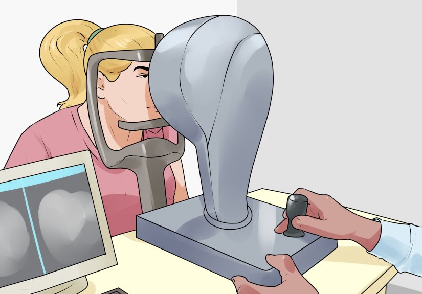
Step 3: Keratometry (measuring corneal curvature)
Keratometry measures the curvature of the corneathe clear front “window” of the eye. Because the cornea provides a large chunk of your eye’s
focusing power, even small shape differences can create astigmatism. Keratometry is especially important for:
- Confirming corneal astigmatism
- Contact lens fitting (especially toric lenses)
- Pre-surgical planning (like cataract or refractive surgery workups)
The test is typically quick and non-contact. You stare at a target while the instrument analyzes reflections to estimate corneal steepness
in different directions. If one meridian is steeper than the other, that difference is corneal astigmatismmeasured in diopters.
Step 4: Corneal topography or tomography (the “map your cornea” upgrade)
If keratometry is a couple of measurements, topography is the whole landscapelike switching from “two weather stations” to “a weather satellite.”
It creates a color-coded map of the cornea’s surface shape. This helps your doctor:
- Measure astigmatism more precisely (including irregular patterns)
- Detect corneal conditions that can distort measurements
- Plan contact lenses and surgical options with better accuracy
Topography is particularly valuable when astigmatism is irregular (not neatly symmetrical), when vision doesn’t match basic
refraction results, or when surgery is on the table. It’s also a big deal if your doctor wants to rule out corneal thinning or shape disorders.
Step 5: Retinoscopy (an objective “light reflex” measurement)
Retinoscopy is a classic technique where the doctor shines a light into your eye and watches how the reflection (“reflex”) moves across your retina.
By introducing lenses and observing changes, they can objectively estimate your refractive errorincluding astigmatism.
It’s especially helpful for kids, patients who can’t reliably answer “1 or 2?”, or situations where the doctor wants an independent check
against the autorefractor and subjective refraction.
Step 6: Subjective refraction (the “1 or 2?” main event)
This is where your prescription becomes your prescription. Using a phoropter (the big lens mask) and a vision chart, the doctor refines
the numbers based on your responses. This step measures astigmatism by dialing in:
- Cylinder power: how much correction you need
- Axis: the best direction for that cylinder
The key tool here is often a Jackson cross cylinder (JCC) technique. In plain English, the doctor flips a special lens back and forth
to compare tiny changes in cylinder and axis. You choose which option looks clearer. This “flip test” is how you go from “pretty good” to “sharp.”
Pro tip: if two choices look the same, it’s okay to say so. Guessing confidently is not a superpower here.
(It’s more like speed-running a video game level you’ve never played.)
Step 7: Slit-lamp and dilated exam (because health matters, too)
Measuring astigmatism isn’t only about numbers. Your doctor also checks eye health: cornea, lens, retina, and more.
A dilated exam can help identify conditions that may affect vision and refraction stability. This doesn’t “measure astigmatism” directly,
but it helps explain why measurements might be changing or why symptoms don’t match the basic prescription.
Regular vs. Irregular Astigmatism: Why the “Shape” Question Matters
Regular astigmatism is the well-behaved kind: the cornea has two principal meridians that are neatly perpendicular.
Glasses and standard toric contacts typically correct it well.
Irregular astigmatism is the rebellious kind: the curvature varies in complex ways, and “one cylinder + one axis” may not fully capture it.
That’s where corneal mapping (topography/tomography) and specialty contact lens testing can become essential.
In some cases, doctors also use a “rigid lens trial” or over-refraction to see how much of the blur is corneal-surface related.
How Doctors Decide the Final Astigmatism Numbers
Here’s the behind-the-scenes logic: your doctor compares multiple data points and looks for agreement.
The final astigmatism measurement usually reflects a balance of:
- Objective estimates (autorefractor, retinoscopy)
- Corneal shape data (keratometry, topography/tomography)
- Your best vision (subjective refraction with JCC)
If everything matches nicely, it’s straightforward. If not, your doctor investigates the “why.”
For example, if corneal topography shows significant corneal astigmatism but subjective refraction shows less cylinder,
the lens inside the eye might be compensatingor the tear film may be affecting surface measurements.
In other cases, the opposite happens, especially if internal (lenticular) astigmatism is contributing.
How to Read Astigmatism on Your Prescription (Without Crying)
Most glasses prescriptions list astigmatism like this:
SPH -2.00 CYL -1.25 Axis 180
- SPH -2.00: nearsighted by 2 diopters
- CYL -1.25: 1.25 diopters of astigmatism
- Axis 180: the cylinder is oriented at 180 degrees (a horizontal axis reference)
You might also see prescriptions written in plus-cylinder form, especially in some clinical settings.
Different format, same optical realityyour optician can translate between them.
What counts as “mild” astigmatism?
There’s no universal cutoff, but many people notice symptoms once cylinder gets to around 0.75D or higherespecially at night
or with lots of screen time. Some people have higher astigmatism and barely notice; others have small amounts and feel like headlights are
doing interpretive dance.
Common Factors That Can Throw Off Astigmatism Measurements
Astigmatism measurement is precise, but eyes are living tissue, not camera lenses. A few common culprits can skew results:
- Dry eye or unstable tear film: can distort corneal readings and cause fluctuating blur
- Contact lens wear: especially rigid lenses, which can temporarily reshape the cornea
- Accommodation: focusing effort can alter refraction, especially in younger patients
- Inconsistent responses: fatigue, rushing, or “I’m just guessing” mode during subjective testing
- Pupil size and lighting: can change how aberrations affect clarity
If your results seem inconsistent, doctors may repeat measurements, use drops to relax focusing (cycloplegia in certain cases),
treat dry eye first, or re-check after you’ve been out of contacts for an appropriate period.
Special Scenarios: When Measuring Astigmatism Gets Extra Important
1) Kids and nonverbal patients
Objective methods like retinoscopy become crucial when someone can’t reliably answer subjective questions.
Accurate astigmatism measurement in childhood can matter for visual development and reducing amblyopia risk.
2) Contact lens fittings (especially toric lenses)
Toric contact lenses correct astigmatism, but they must align properly on the eye. Keratometry and corneal mapping help predict fit,
while over-refraction confirms real-world clarity once the lens settles.
In other words: the “numbers” are step one; the “how it behaves on your eye” is step two.
3) Pre-op planning for refractive surgery or cataract surgery
Surgical planning often requires very accurate corneal measurements and consistent refraction data.
Corneal topography/tomography is commonly used to map astigmatism and screen for irregularities.
For cataract surgery, astigmatism measurement can influence whether toric intraocular lenses or other strategies are appropriate.
Conclusion: The Best Astigmatism Measurement Is a Team Effort
Measuring astigmatism isn’t a single testit’s a smart combination of objective instruments, corneal shape mapping, and the most important tool of all:
your real-world visual clarity. The autorefractor and keratometry provide strong clues. Retinoscopy backs it up.
Corneal topography adds detail when the situation is complex. And subjective refraction refines everything into the prescription you’ll actually wear.
If you ever feel like your exam is “a lot of steps,” that’s a good sign. It means your doctor is cross-checking data to make sure your final cylinder
and axis aren’t just accuratethey’re useful for your daily life. Your eyes deserve more than a guess. They deserve a well-edited final draft.
Real-World Experiences: What Measuring Astigmatism Actually Feels Like (and How to Nail It)
Let’s talk about the part nobody warns you about: the emotional journey of being asked “Which is better… one… or two?” approximately
47 times in a row. If you’ve ever left an eye exam thinking, “I’m pretty sure I accidentally picked a new personality,” you’re not alone.
Measuring astigmatism is precisebut the patient experience can be oddly human.
Experience #1: The letters look the same.
This happens constantly during cylinder/axis refinement. The differences can be subtlelike comparing two identical photos where one is 2% sharper.
The best move is honesty: say “about the same” or “they’re both blurry.” Clinicians can interpret that response and adjust strategy
(bigger steps, re-checking sphere, or revisiting the starting point). Confident guessing doesn’t help. It’s not a game show;
there is no prize wheel behind the phoropter.
Experience #2: Night driving complaints that don’t match “mild” numbers.
Many people first notice astigmatism as halos, glare, starbursts, or smeared headlightsespecially at night.
Even smaller amounts of cylinder can feel bigger in low light because pupils dilate, letting more optical imperfections contribute.
Patients often describe it as “everything has a ghost outline.” Bringing specific examples helps: “Street signs bloom,” “headlights starburst,”
“I squint to sharpen things.” Those details can guide extra testing (like corneal mapping or careful dry-eye assessment).
Experience #3: The dry-eye plot twist.
Dry eye is the sneakiest saboteur of consistent astigmatism readings. The tear film is the first refractive surface of the eye.
If it’s uneven, the front of the eye behaves like a constantly changing lensmeaning your cylinder and axis can wobble.
A common real-world scenario: the first measurement says one thing, you blink a lot, and the next reading shifts.
People sometimes feel “my vision changes every minute,” especially with screens, heating/AC, or contact lens wear.
If your clinician recommends treating dryness before finalizing a prescription, it’s not a stalling tacticit’s quality control.
Experience #4: Contact lenses and the “my cornea needs a nap” effect.
Some contact lensesparticularly rigid lensescan temporarily change corneal shape.
If you’re being measured for glasses, surgery planning, or specialty lenses, you may be asked to stop wearing contacts for a period beforehand.
Patients sometimes find this annoying (“But I need to see to live!”), so a practical workaround is scheduling:
use older glasses for a few days, plan the exam when you can be a little blurry, and communicate your lens-wear history clearly.
The payoff is more reliable keratometry/topography and a prescription that doesn’t feel “off” a week later.
Experience #5: How to be a “great patient” during astigmatism testing.
You don’t need perfect answersyou need consistent ones. Try these:
- Don’t squint. Squinting can temporarily sharpen vision and confuse the refinement process.
- Pick the clearest overall, not the darkest. Some lenses make letters look bolder but not sharper.
- Ask for a repeat if you’re unsure. “Can I see that again?” is totally valid.
- Describe what you see. “Sharper edges,” “less shadow,” “less smear,” or “same” gives useful data.
- Tell them about your real life. Computer-heavy job? Night driving? Sports? That context matters.
The most reassuring takeaway from real exam chairs: measuring astigmatism is a collaboration. The instruments provide the science;
your feedback provides the reality check. And when those line up, your prescription stops feeling like a random number soup and starts feeling like:
“Oh. This is what crisp looks like.”